Заключение
Несмотря на различия в механизмах генеза отставленного когнитивного дефицита, наблюдаемого после пренатальной патологии, и при развитии такого нейродегенеративного заболевания, как болезнь Альцгеймера, обе эти патологии имеют общие особенности.
Как при пренатальной гипоксии, так и на ранних стадиях МКС происходит дисрегуляция синапс-ассоциированных белков, сопровождающаяся комплексными нарушениями межнейронного взаимодействия, в первую очередь снижением пластичности и адаптивного потенциала кортикальных отделов головного мозга. Это провоцирует у таких животных когнитивные дисфункции на взрослой стадии развития. Повышение активности каспаз в раннем постнатальном онтогенезе животных, перенесших пренатальную гипоксию, может вызывать деградацию синапс-ассоциированных белков и дегенерацию синаптических структур, вызывая когнитивный дефицит на взрослой стадии развития. Изменение уровня активности ферментов, участвующих в образовании и катаболизме Ар, являющегося основным токсическим агентом, приводящим к БА, также создает предпосыл-Таблица
Аналаз характеристики поведения, структурно-функциональных и биохимических изменений в структурах мозга экспериментальных животных
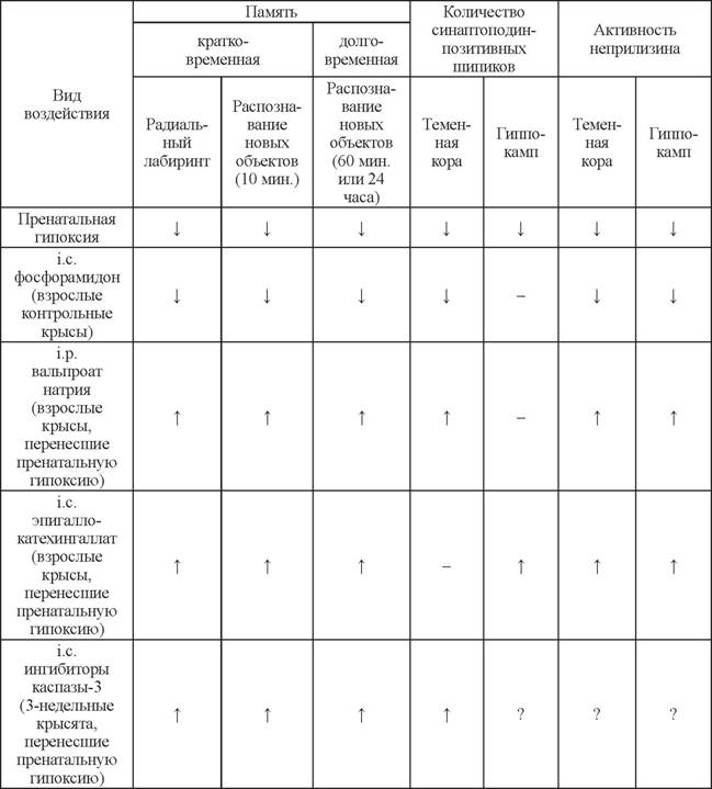
T - улучшение или увеличение по сравнению с животными, перенесшими пренатальную гипоксию,
4 - ухудшение или уменьшение по сравнению с контролем,-------- нет статистически достоверных
изменений, ? - нет данных.
ки к его накоплению в процессе старения мозга. Эти же изменения наблюдаются после пренатальной гипоксии на более ранних стадиях развития. Накопленные к настоящему времени результаты исследований, проводимых в нашей лаборатории (табл.), и данные литературы подтверждают, что модель пренатальной гипоксии является адекватным инструментом для изучения молекулярных механизмов ранних этапов развития когнитивных дисфункций и удобным объектом для разработки и тестирования лекарственных препаратов.
Литература
Васильев Д.С., Туманова Н.Л., Журавин И.А. Изменение нервной ткани новой коры в онтогенезе крыс после гипоксии на разных сроках онтогенеза // Журнал эволюц. биохим. физиол. 2008. Т 44. № 3. С. 258-266.
Васильев Д.С., Туманова Н.Л., Журавин И.А. Изменение межклеточного взаимодействия в кортикальных отделах мозга крыс после пренатальной гипоксии // Материалы XXII Съезда Физиологического общества им. И.П. Павлова, г. Волгоград, 16-20 сентября. 2013. С. 531-532.
Дубровская Н.М., Журавин И.А. Онтогенетические особенности поведения крыс, перенесших гипоксию на 14-е или 18-е сутки эмбриогенеза // ЖВНД. 2008. Т. 58. № 5. С. 616-625.
Дубровская Н.М., Наливаева Н.Н., Плеснева С.А. и др. Изменение активности амилоид-дегради- рующих металлопептидаз приводит к нарушению памяти у крыс // ЖВНД. 2009. Т 59. № 5. С. 630-638.
Журавин И.А., Дубровская Н.М., Туманова Н.Л. Постнатальное физиологическое развитие крыс после острой пренатальной гипоксии // Российский физиол. журнал им. И.М. Сеченова. 2003. Т 89. № 5. С. 522-532.
Журавин И.А., Туманова Н.Л., Озирская Е.В. и др. Формирование структурной и ультраструктурной организации стриатума в раннем постнатальном онтогенезе крыс при изменении условий их эмбрионального развития // Морфология. 2005. Т 127. № 2. С. 31-36.
Журавин И.А., Туманова Н.Л., Озирская Е.В. и др. Формирование структурной и ультраструктурной организации стриатума в постнатальном онтогенезе крыс при изменении условий их эмбрионального развития // Журнал эвол. биох. физиол. 2007а. Т. 43. № 2. С. 199-208.
Журавин И.А., ДубровскаяН.М., КочкинаЕ.Г и др. Исследование действия гипоксии на развитие функций мозга и метаболизм амилоидного пептида с целью разработки средств ранней диагностики и профилактики болезни Альцгеймера // Технол. жив. сист. 2007б. Т. 4. № 5-6. С. 109-122.
Журавин И.А., Туманова Н.Л., Васильев Д.С. Структурные изменения нервной ткани гиппокампа в онтогенезе крыс после пренатальной гипоксии // Журнал эвол. биох. физиол.
2009а. Т. 45. № 1. С. 138-140.Журавин И.А., Туманова Н.Л., Васильев Д.С. Изменение адаптивных механизмов мозга в онтогенезе крыс, перенесших пренатальную гипоксию // Докл. РАН. 2009б. Т 425. № 1. С. 123-125.
Журавин И.А., Дубровская Н.М., Васильев Д.С. и др. Эпигенетическая и фармакологическая регуляция амилоид-деградирующего фермента неприлизина приводит к изменению когнитивных функций млекопитающих // Докл. РАН. 2011. T. 438. № 6. C. 838-841.
Захаров В.В., Яхно Н.Н. Когнитивные расстройства в пожилом и старческом возрасте. Методическое пособие для врачей. М., 2005.
Кочкина Е.Г., Плеснева С.А., Журавин И.А. и др. Влияние гипоксии на активность холинэстераз в сенсомоторной коре мозга крыс // Журнал эвол. биох. физиол. 2015. Т. 51. № 1. В печати.
Лавренова С.М., Наливаева Н.Н., Журавин И.А. Активность ацетилхолинэстеразы сенсомоторной коры в раннем онтогенезе крыс, перенесших пренатальную гипоксию // Журнал эвол. биох. фи- зиол. 2003. Т 39. C. 154-159.
Отеллин В.А., Хожай Л.И., Гилеpович Е.Г. и др. Повреждающие воздействия в критическиепериоды- пренатального онтогенеза как фактор, модифицирующийструктурноеразвитие головного мозга и поведенческие реакции после рождения // Вестник РАМН. 2002. Т. 12. С. 32-35.
Algan O., Rakic P. Radiation-induced, Іатіпа^ресШс deletion of neurons in the primate visual cortex // J. Сотр. Neurol. 1997. Vol. 381. P. 1096-9861.
Ang E.S. Jr., Gluncic V., Duque A. et al. Prenatal exposure to ultrasound waves тра^ neuronal migration in mice // Proc. Natl. Acad. Sci. USA. 2006. Vol. 103. P. 12903-12910.
Arendt T. Synaptic plasticity and cell cycle activation in neurons are alternative effector pathways: the ‘Dr. Jekyll and Mr. Hyde concept’ of Alzheimer’s disease or the yin and yang of neuroplasticity // Prog. Neurobiol. 2003. Vol. 71. P. 83-248.
Asanuma K., Kim K., Oh J. et al. Synaptopodin regulates the actin-bundling activity of a-actinin in an isoform-specific manner // J. Clin. Invest. 2005. Vol. 115. P. 1188-1198.
Beckett C., Bagrova D.I., Belyaev N.D., TurnerA.J., Nalivaeva N.N. Caspase cleavage of the amyloid precursor protein intracellular domain (AICD) decreases neprilysin expression and increases Ap in hypoxia // J. Neurochem. 2012. Vol. 123. P. 101.
Belayev N.D., Nalivaeva N.N., Makova N.Z. et al. Neprilysin gene expression requires binding of the amyloid precursor protein intracellular domain to its promoter: implications for Alzheimer disease // EMBO Rep. 2009. Vol. 10. P. 94-100.
Belyaev N.D., Kellett K.A., Beckett C. et al. The transcriptionally active amyloid precursor protein (APP) intracellular domain is preferentially produced from the 695 isoform of APP in a P-secretase-dependent pathway // J. Biol. Chem. 2010. Vol. 285. P. 41443-41454.
Broughton B.R., Reutens D.C., Sobey C.G. Apoptotic mechanisms after cerebral ischemia // Stroke. 2009. Vol. 40. P. 331-339.
Cavanaugh S.E., Pippin J.J., Barnard N.D. Animal models of Alzheimer disease: historical pitfalls and a path forward // ALTEX. 2014. doi:http://dx.doi.org/10.14573/altex.1310071.
Chen Y., Rex C.S., Rice C.J. et al. Correlated memory defects and hippocampal dendritic spine loss after acute stress involve corticotropin-releasing hormone signaling // Proc. Natl. Acad. Sci. USA. 2010. Vol. 107. P. 13123-13128.
Cheng I.H., Scearce-Levie K., Legleiter J. et al. Accelerating amyloid-beta fibrillization reduces oligomer levels and functional deficits in Alzheimer disease mouse models // J. Biol. Chem. 2007. Vol. 282. P. 23818-23828.
Chowdhury I.,Tharakan B., Bhat G.K. Caspases - an update // Comp. Biochem. Physiol. B Biochem. Mol. Biol. 2008. Vol. 151. P. 10-27.
Cohen R.M., Rezai-Zadeh K., Weitz T.M. et al. A transgenic Alzheimer rat with plaques, tau pathology, behavioral impairment, oligomericabeta, and Frank neuronal loss // J. Neurosci. 2013. Vol. 33. P. 6245-6256.
DAmelioM, Cavallucci V., CecconiF. Neuronal caspase-3 signaling: not only cell death // Cell. Death. Differ. 2010. Vol.
17. P. 1104-1114.DAmelioM., Cavallucci V., Middei S.et al. Caspase-3 triggers early synaptic dysfunction in a mouse model of Alzheimer’s disease // Nat. Neurosci. 2011. Vol. 14. P. 69-76.
De la Monte S.M., Wands J.R. Review of insulin and insulin-like growth factor expression, signaling, and malfunction in the central nervous system: relevance to Alzheimer’s disease // J. Alzheimers Dis. 2005. Vol. 7. P. 45-61.
Delivoria-Papadopoulos M., Ashraf Q.M., Ara J. et al. Nuclear mechanisms of hypoxic cerebral injury in the newborn: the role of caspases // Semin. Perinatol. 2008. Vol. 32. P. 334-43.
Deller T., Korte M., Chabanis S. et al. Synaptopodin-deficient mice lack a spine apparatus and show deficits in synaptic plasticity // PNAS. 2003. Vol. 100. P. 10494-10499.
Deller T., Bas Orth C., Vlachos A. et al. Plasticity of synaptopodin and the spine apparatus organelle in the rat fascia dentata following entorhinal cortex lesion // J. Compar. Neurol. 2006. Vol. 499. P. 471-484.
Del Rio J.A., Martinez A., Auladell C. et al. Developmental history of the subplate and developing white matter in the murine neocortex. Neuronal organization and relationship with the main afferent systems at embryonic and perinatal stages // Cerebral Cortex. 2000. Vol. 10. P. 784-801.
Del Valle J., Duran-Vilaregut J., Manich G. et al. Early amyloid accumulation in the hippocampus of SAMP8 mice // J. Alzheimers Dis. 2010. Vol. 19. P. 1303-1315.
Dubrovskaya N.M., Nalivaeva N.N., Vasilev et al. Mechanisms of short-term working memory deficit // Short-Term Memory: New Research. Eds. G. Kalivas, S.F. Petralia. NY: Nova Science Publishers Inc., 2012. Ch. 6. P. 155-173.
Dumas J.A., Newhouse P.A. The cholinergic hypothesis of cognitive aging revisited again: cholinergic functional compensation // Pharmacol. Biochem. Behav. 2011. Vol. 99. P. 254-261.
Fan T.J., Han L.H., Cong R.S. et al. Caspase family proteases and apoptosis // Acta. Biochim. Biophys. Sin. (Shanghai). 2005. Vol.
37. P. 719-727.Flood J.F., Cherkin A. Scopolamine effects on memory retention in mice: a model of dementia? // Behav. Neural. Biol. 1986.Vol. 45. P. 169-184.
Gerstein M., Huleihel M., Mane R. et al. Remodeling of hippocampal GABAergic system in adult offspring after maternal hypoxia and magnesium sulfate load: immunohistochemical study // Exp. Neurol. 2005. Vol. 196. P. 18-29.
Golan M.H., Mane R., Molczadzki G. et al. Impaired migration signaling in the hippocampus following prenatal hypoxia // Neuropharmacology. 2009. Vol. 57. P. 511-522.
Gomez-Isla T, Price J.L., McKeel D.W. Jr. et al. Profound loss of layer II entorhinal cortex neurons occurs in very mild Alzheimer’s disease // J. Neurosci. 1996. Vol. 16. P. 4491-4500.
Greig N.H., Lahiri D.K., Sambamurti K. Butyrylcholinesterase: an important new target in Alzheimer’s disease therapy // Int. Psychogeriatr. 2002. Vol. 14. Suppl. 1. P. 77-91.
Grunblatt E., Hoyer S., Riederer P. Gene expression profile in streptozotocin rat model for sporadic Alzheimer’s disease // J. Neural. Transm. 2004. Vol. 111. P. 367-386.
Guan H., Liu Y., Daily A. et al. Peripherally expressed neprilysin reduces brain amyloid burden: a novel approach for treating Alzheimer’s disease // J. Neurosci. Res. 2009. Vol. 87. P. 1462-1473.
Gulyaeva N.V Non-apoptotic functions of caspase-3 in nervous tissue // Biochemistry (Mosc). 2003. Vol. 68. P. 1171-1180.
HallidayA.C., GreenfieldS.A. From protein to peptides: a spectrum of non-hydrolytic functions of acetylcholinesterase // Protein Pept. Lett. 2012. Vol. 19. P. 165-72.
Hardy J. The amyloid hypothesis for Alzheimer’s disease: a critical reappraisal // J. Neurochem. 2009. Vol. 110. P. 1129-1134.
Hardy J., Selkoe D.J. The amyloid hypothesis of Alzheimer’s disease: progress and problems on the road to therapeutics // Science. 2002. Vol. 297. P. 353-356.
Hetz C., GlimcherL. The daily job of night killers: alternative roles of the BCL-2 family in organelle physiology // Trends Cell. Biol. 2008. Vol. 18. P. 38-44.
Hwang, D.Y., Cho, J.S., Oh, J.H. et al. Early changes in behavior deficits, amyloid beta-42 deposits and MAPK activation in doubly transgenic mice co-expressing NSE-controlled human mutant PS2 and AP- Psw // Cell. Mol. Neurobiol. 2005. Vol. 25. P. 881-898.
Itoh M., Li S., Ohta K. et al. Cayman ataxia-related protein is a presynapse-specific caspase-3 substrate // Neurochem. Res. 2011.Vol. 36. P. 1304-1313.
Jiao S., Li Z. Nonapoptotic function of BAD and BAX in long-term depression of synaptic transmission // Neuron. 2011. Vol. 70. P. 758-772.
Kudryashov I.E., Yakovlev A.A., Kudryashova I. et al. Footshock stress alters early postnatal development of electrophysiological responses and caspase-3 activity in rat hippocampus // Neurosci. Lett. 2002. Vol. 332. P. 95-98.
Kudryashova I.V., Stepanichev M.Y., Gulyaeva N.V. Natural activation of caspase-3 is required for the development of operant behavior in postnatal ontogenesis // Neurosci. Behav. Physiol. 2009a. Vol. 39. P. 65-72.
Kudryashova I.V., Onufriev M.V., Kudryashov I.E. et al. Caspase-3 activity in hippocampal slices reflects changes in synaptic plasticity // Neurosci. Behav. Physiol. 2009b. Vol. 39. P. 13-20.
Lema Tome C.M., Nottingham C.U., Smith C.M. et al. Neonatal exposure to MK801 induces structural reorganization of the central nervous system // Neuroreport. 2006. Vol. 17. P. 779-783.
Lesne S., Koh M.T., KotilinekL. et al. A specific amyloid-beta protein assembly in the brain impairs memory // Nature. 2006. Vol. 440. P. 352-357.
Louzoun-Kaplan V., Zuckerman M., Perez-Polo J.R. et al. Prenatal hypoxia down regulates the GABA pathway in newborn mice cerebral cortex; partial protection by MgSO4 // Int. J. Dev. Neurosci. 2008. Vol. 26. P. 77-85.
Martin K.C., BaradM., KandelE.R. Local protein synthesis and its role in synapse-specific plasticity // Currr. Opin. Neurobiol. 2000. Vol. 10. P. 587-592.
McDonald M.P., Overmier J.B. Present imperfect: a critical review of animal models of the mnemonic impairments in Alzheimer’s disease // Neurosci. Biobehav. Rev. 1998. Vol. 22. P. 99-120.
MelzigM.F., Janka M. Enhancement of neutral endopeptidase activity in SK-N-SH cells by green tea extract // Phytomedicine. 2003. Vol. 10. P. 494-498.
Morrissette D.A., Parachikova A., Green K.N. et al. Relevance of transgenic mouse models to human Alzheimer disease // J. Biol. Chem. 2009. Vol. 284. P. 6033-6037.
Mundel P., Heid H.W., Mundel T.M. et al. Synaptopodin: an actin-associated protein in telencephalic dendrites and renal podocytes // J. Cell. Biol. 1997. Vol. 139. P. 193-204.
Nalivaeva N.N., Turner A.J. Does acetylcholinesterase secretion involve an ADAMs-like metallosecretase? // Lett. Pept. Sci. 1999. Vol. 6. P. 343-348.
Nalivaeva N.N., Turner A.J. The amyloid precursor protein: a biochemical enigma in brain development, function and disease // FEBS Lett. 2013. Vol. 587. P. 2046-2054.
Nalivaeva N.N., Beckett C., Belyaev N.D., Turner A.J. Are amyloid-degrading enzymes viable therapeutic targets in Alzheimer’s disease? // J. Neurochem. 1992. Vol. 120. Suppl. 1. P. 167-185.
Nalivaeva N.N., Fisk L. CanetAviles, R.M. et al. Effects of prenatal hypoxia on expression of amyloid precursor protein and metallopeptidases in the rat brain // Lett. Pept. Sci. 2003. Vol. 10. P. 455-462.
Nalivaeva N.N., Fisk L., Kochkina E.G. et al. Effect of hypoxia/ischemia and hypoxic preconditioning/re- perfusion on expression of some amyloid-degrading enzymes // Ann. NY Acad. Sci. 2005. Vol. 1035. P. 21-33.
Nalivaeva N.N., Belyaev N.D., Lewis D.I. et al. Effect of sodium valproate administration on brain neprilysin expression and memory in rats // J. Mol. Neurosci. 2012a. Vol. 46. P. 569-577.
Nalivaeva N.N., Belyaev N.D., Lewis D.I. et al. Effect of sodium valproate administration on brain neprilysin expression and memory in rats // J. Mol. Neurosci. 2012b. Vol. 46. P. 569-577.
Newman T, Sinadinos C., Johnston A. et al. Using Drosophila models of neurodegenerative diseases for drug discovery // Expert Opin. Drug. Discov. 2011. Vol. 6. P. 129-140.
Nyakas C, Buwalda B., Luiten P.G.M. Hypoxia and brain development // Prog. in Neurobiol. 1996. Vol. 49. P. 1-51.
Oddo S., Caccamo A., Shepherd J.D. et al. Triple-transgenic model of Alzheimer’s disease with plaques and tangles: intracellular Abeta and synaptic dysfunction // Neuron. 2003. Vol. 39. P. 409-421.
Okubo-Suzuki R., Okada D., Sekiguchi M. et al. Synaptopodin maintains the neural activity-dependent enlargement of dendritic spines in hippocampal neurons // J. Mol. Cell. Neurosci. 2008. Vol. 38. P. 266-276.
Oomman S., Finckbone V., Dertien J. et al. Active caspase-3 expression during postnatal development of rat cerebellum is not systematically or consistently associated with apoptosis // J. Comp. Neurol. 2004. Vol. 476. P. 154-73.
Pardossi-Piquard R., Petit A., Kawarai T. Presenilin-dependent transcriptional control of the AP-degrading enzyme neprilysin by intracellular domains of PAPP and APLP // Neuron. 2005. Vol. 46. P 541-554.
Pepeu G. Mild cognitive impairment: animal models // Dialogues Clin. Neurosci. 2004. Vol. 6. P. 369-377.
Pepeu G., Casamenti F., Pedata F. et al. Are the neurochemical and behavioral changes induced by lesions of the nucleus basalis in the rat a model of Alzheimer’s disease? // Prog. Neuropsychopharmacol. Biol. Psychiatry. 1986. Vol. 10. P 541-551.
Prescott S.A., Chase R. Sites of plasticity in the neural circuit mediating tentacle withdrawal in the snail Helix aspersa: implications for behavioral change and learning kinetics // Learn. Mem. 1999. Vol. 6. P. 363-380.
Prickaerts J., Fahrig T., BloklandA. Cognitive performance and biochemical markers in septum, hippocampus and striatum of rats after an i.c.v. injection of streptozotocin: a correlation analysis // Behav. Brain Res. 1999. Vol. 102. P 73-88.
RakicP. Defects of neuronal migration and pathogenesis of cortical malformations // Prog. Brain.Res. 1988. Vol. 73. P 15-37.
Rice D., Baron S. Critical periods of vulnerability for the developing nervous system: evidence from humans and animal models // Environ. Health Perspect. 2000. Vol. 108. P 511-533.
Roth K.A., D’Sa C. Apoptosis and brain development // Ment. Retard. Dev. Disabil. Res. Rev. 2001.Vol. 7. P. 261-266.
Salamone J.D., Lalies M.D., Channell S.L., Iversen S.D. Behavioural and pharmacological characterization of the mouth movements induced by muscarinic agonists in the rat // Psychopharmacology (Berl). 1986. Vol. 88. P. 467-471.
Salkovic-Petrisic M., Tribl F., Schmidt M. et al. Alzheimer-like changes in protein kinase B and glycogen synthase kinase-3 in rat frontal cortex and hippocampus after damage to the insulin signalling pathway // J. Neurochem. 2006. Vol. 96. P 1005-1015.
Salkovic-Petrisic M., Knezovic A., Hoyer S., Riederer P. What have we learned from the streptozotocin- induced animal model of sporadic Alzheimer’s disease, about the therapeutic strategies in Alzheimer’s research // J. Neural. Transm. 2013. Vol. 120. P. 233-252.
Sanchez R.M., Ribak C.E., Shapiro L.A. Synaptic connections of hilar basal dendrites of dentate granule cells in a neonatal hypoxia model of epilepsy // Epilepsia. 2012. Vol. 53. Suppl. 1. P. 98-108.
Santos H.R., Cintra W.M., Aracava Y. et al. Spine density and dendritic branching pattern of hippocampal CA1 pyramidal neurons in neonatal rats chronically exposed to the organophosphate paraoxon // Neurotoxicology. 2004. Vol. 25. P. 481-494.
Saura C.A., Chen G., Malkani S. et al. Conditional inactivation of presenilin 1 prevents amyloid accumulation and temporarily rescues contextual and spatial working memory impairments in amyloid precursor protein transgenic mice // J. Neurosci. 2005. Vol. 25. P. 6755-6764.
Schliebs R., Arendt T. The cholinergic system in aging and neuronal degeneration // Behav. Brain. Res. 2011. Vol. 221. P. 555-563.
Segal M. Dendritic spines, synaptic plasticity and neuronal survival: activity shapes dendritic spines to enhance neuronal viability // Eur. J. Neurosci. 2010. Vol. 31. P. 2178-2184.
Shapiro L.A., Ribak C.E. Integration of newly born dentate granule cells into adult brains: hypotheses based on normal and epileptic rodents // Brain. Res. Rev. 2005. Vol. 48. P. 43-56.
SchaefferE.L., FigueiroM., Gattaz W. F. Insights into Alzheimer disease pathogenesis from studies in transgenic animal models // Clinics (Sao Paulo). 2011. Vol. 66. Suppl 1. P. 45-54.
Snigdha S., Smith E.D., Prieto G.A. et al. Caspase-3 activation as a bifurcation point between plasticity and cell death // Neurosci. Bull. 2012. Vol. 28. P 14-24.
Sousa N., Lukoyanov N. V., Madeira M.D. et al. Reorganization of the morphology of hippocampal neurites and synapses after stress-induced damage correlates with behavioral improvement // Neurosci. 2000. Vol. 97. P. 253-66.
Sultana R., Banks W.A., Butterfield D.A. Decreased levels of PSD95 and two associated proteins and increased levels of BCl2 and caspase3 in hippocampus from subjects with amnestic mild cognitive impairment: Insights into their potential roles for loss of synapses and memory, accumulation of Ap, and neurodegeneration in a prodromal stage of Alzheimer’s disease // J. Neurosci. Res. 2010. Vol. 88. P. 469-77.
Takeda S., Sato N., Niisato K. et al., Validation of AP1-40 administration into mouse cerebroventricles as an animal model for Alzheimer disease // Brain. Res. 2009. Vol. 1280. P. 137-147.
Vasilev D.S.,Tumanova N.L., Dubrovskaya N.M. et al. Prenatal hypoxia affects neuronal generation and migration with selective impact on different cell populations in rat neocortex // FENS Forum for Neuroscience. Barselona, 2012. Abstract Number 2225.
Walsh D.M., Klyubin I., Fadeeva J.V. et al. Amyloid-P oligomers: their production, toxicity and therapeutic inhibition // Biochem. Soc. Trans. 2002. Vol. 30. P. 552-557.
Weinreb O., Mandel S., Amit T. et al. Neurological mechanisms of greentea polyphenols in Alzheimer’s and Parkinson’s diseases // J. Nutr. Biochem. 2004. Vol. 15. P. 506-16.
Wiley R.G., Oeltmann T.N., Lappi D.A. Immunolesioning: selective destruction of neurons using immuno- toxin to rat NGF receptor // Brain. Res. 1991. Vol. 562. P. 149-153.
Zhu Z.J., Wu K.C., Qian Z.M. et al. Drosophila models for studying iron-related neurodegenerative diseases // Sheng Li Xue Bao. 2014. Vol. 66. P 47-54.
Zito K., Scheuss V., Knott G. et al. Rapid functional maturation of nascent dendritic spines // Neuron. 2009. Vol. 61. P 247-258.
Еще по теме Заключение:
- Заключение
- Заключение
- Заключение
- Заключение
- Заключение
- Заключение
- Заключение
- Заключение
- 2.5. Рациональное трудоустройство больных по заключению КЭК
- Патоморфологическое заключение
- Вместо заключения
- Параграф пятый. Об общих заключениях по действиям мозга
- Механизмы заключения договоров
- Заключение: от понимания к действию